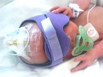
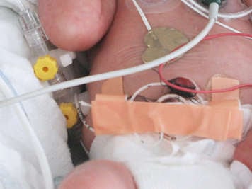
There are several types of lines and catheters that can be used for vascular access on a premature baby. The vascular devices are small catheters or tubes inserted into either a vein or an artery.
Central venous lines
Central type catheters, also called central lines, are inserted into a vein in one part of the body and the tube is then guided within the vein to a point near the heart. The choice of vascular access that is used depends on the gestational age and condition of the baby. In general, arterial lines, which provide access to the arteries, are used to draw blood or to monitor blood pressure. Venous lines, which provide access to the veins, are used to deliver nutrition, fluids, and medications, though they can also be used to take blood samples.
The catheters are fed through the vascular system to a point near the heart for several reasons:
- A premature baby’s veins can be fragile, so placing the tip near one of the larger veins means that it can last longer, is less likely to become dislodged, and is less likely to cause damage.
- Blood flow is strongest near the heart, which means the flow of medicine, nutrition, or fluids through the catheter will be stronger and more constant. If it is delivered at a point near the heart, medicine will diffuse throughout the body more quickly. This allows for higher concentrations of medicines to be delivered through the lines without irritating the veins. For example, high sugar solutions can be irritating to the veins and so they are usually only administered intravenously through a central line and not through a peripheral intravenous (PIV) line, described in the next section.
Depending on the premature baby’s condition, several lines may be needed at the same time. For example, one line might be used for nutrition while another separate line delivers medicine. Babies requiring several separate treatments are more likely to have more than one line. Some catheters have two or more separate tubes, or lumina, within one tube, rather like two trains running on different tracks through a tunnel. This may allow separate treatments, such as delivery of two medications, to occur at the same time.
All methods of vascular access require regular maintenance and monitoring. For example, heparin, an agent that prevents blood clotting, is used in some hospitals to keep the lines of the catheter from clogging up. Additionally, all types of vascular access can cause complications, most of which are rare but can be severe.

Peripheral intravenous lines
Peripheral intravenous (PIV) lines are catheters placed in veins in the hands, feet, or scalp. PIV lines can be inserted and left in place for a few days, and are generally used for delivery of nutrition or medication, and hydrating fluids. These lines are not threaded as deep into the body and usually last for hours to a few days; they are not the best option for babies who may need longer term IV therapy.
Umbilical venous catheters

An umbilical venous catheter (UVC) is placed into the cardiovascular system through the remaining “tail” of a cut umbilical cord. Generally, they are the preferred method of venous access in the Neonatal Intensive Care Unit (NICU), however, the line can only be inserted into a recently newborn baby. After about two weeks out of the womb, the umbilical cord and the veins leading from it to the body naturally obliterate and disappear. UVC lines can be placed quickly, can remain in place for up to two weeks if necessary, and can accommodate a larger size catheter. The catheter is fed from the umbilical cord to just outside a chamber of the heart called the right atrium.
Peripherally inserted central venous catheters
Peripherally inserted central venous catheters (PICC lines) are placed into a peripheral vein and then fed through the vascular system until the tip of the catheter is in the superior vena cava, which is one of the major veins near the heart. Usually, the threading of the catheter begins in a vein in the arm, leg, or scalp. For newborns having a PICC line inserted in the NICU, a chest X-ray is done to visually ensure correct placement of the tip. Sometimes, babies must go to the Interventional Guided Therapy (IGT) suite to have the PICC inserted. An IGT suite is a room equipped with imaging devices that help doctors insert and thread the line to the right place. Following line insertion in the IGT, the correct placement of the tip is confirmed by fluoroscopy, another type of imaging technology. A PICC line can be left in place for several weeks. If a baby is expected to need an IV for an extended period, a PICC line may be used.