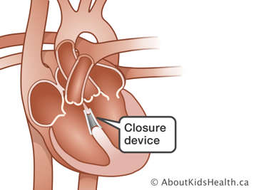
For some heart defects such as patent ductus arteriosus, atrial septal defect, and ventricular septal defect, a small closure device can be inserted into the heart to fill a hole in the heart. In some cases, this is enough to correct the problem.
What does the closure device look like and how does it work?
There are a number of different kinds of closure devices. For example, one device looks like two opened umbrellas when it is in position. The device has spring arms that hold it in the proper place and prevent blood from flowing through the hole. The arms stay closed until it reaches the right position in the hole. It is then opened up like an umbrella. When it comes to repairing a PDA, you may hear this device referred to as a PDA coil.

How is a closure device inserted?
The closure procedure is done in a special cardiac procedure room. The procedure is performed while your child is under a general anaesthetic. This means that your child will be asleep during the procedure.
Not every hole can be closed with a device. Some holes are too big. An echocardiogram taken from the esophagus or stomach tube (a transesophageal echocardiogram) is done to measure the size of the hole and to help place the device. If the echocardiogram shows that the hole is too big to close with the device, your child will be awakened and sent to the recovery room.
If the hole is small enough and in the right position, thin tubes called catheters will be inserted through the blood vessels in the groin at the top of the leg (sometimes both legs are used). The catheter is moved up the blood vessel into the heart and into the affected area of the heart. The closure device is inserted through one catheter, while the other is used to help measure the hole. After the device is in place, the catheters are taken out and the entry sites are covered with a bandage. An echocardiogram might be done to check the placement of the device.
If the device doesn't fit properly after it is opened, it may be taken out and a second attempt made to close the hole. If the device cannot be taken out, which is rare, surgery will be necessary. A more common complication is a small leak through the closure device. Usually this disappears with time. In this case, a second device may be placed over the first to stop the leak completely.