Ventricular septal defect (VSD)

PDF download is not available for Arabic and Urdu languages at this time. Please use the browser print function instead

Learn about ventricular septal defects (VSD). These defects are holes that develop in the heart that force it to overwork, making it get bigger.

Key points

  • With ventricular septal defects (VSD), an excess of blood can travel to the lungs.
  • VSD is usually detected when a doctor hears a heart murmur.
  • Treatment for VSDs depends on the size of the hole and can range from no treatment to open heart surgery.
  • The outlook for children with this condition is very good.

An opening in the wall between the two lower chambers of the heart is known as a ventricular septal defect. A VSD causes blood high in oxygen to leak from the left side of the heart, where the pressure is higher, to the right side. With large holes, this is a problem because too much blood flows to the lungs. This forces the heart to overwork and makes it bigger. There are four types of VSDs, named according to where they occur in the septum: membranous defect, muscular defect, inlet defect, and outlet defect.

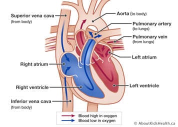
The two images below compare a normal heart to a heart with a ventricular septal defect.

Normal heart Normal heart showing placement of the atria, ventricles, aorta, superior vena cava, inferior vena cava, pulmonary artery and pulmonary veins
The heart has two upper (receiving) and two lower (pumping) chambers. Blood flows into the upper chambers (the right atrium and the left atrium). The lower chambers (the right and left ventricles) pump blood out of the heart. The heart valves open and close to keep blood flowing in the correct direction.
Ventricular septal defect Heart showing pulmonary artery with extra blood flow to lungs, enlarged left ventricle, right ventricle, and a hole between ventricles allowing blood to flow from left to right ventricle.
A ventricular septal defect (VSD) is a hole in the wall dividing the two lower chambers of the heart, the right and left ventricle. This hole allows blood to pass from the left ventricle into the pulmonary artery and then to the lungs. The amount of extra blood flow to the lungs depends on the size of the VSD.

VSD accounts for about 20% of congenital heart defects. About five to 50 children out of 1,000 are born with this defect.

What are the symptoms of ventricular septal defect?

The hole does not usually cause symptoms if it is small and does not allow much blood to pass through. For about 5% to 10% of cases, though, the hole may be large and cause congestive heart failure. A heart murmur may also be present.

These defects are usually identified when a doctor hears a heart murmur. A chest X-ray, electrocardiogram, and echocardiogram or, in rare cases, cardiac catheterization, may be done to diagnose this condition.

How is ventricular septal defect treated?

VSDs occur in a range of sizes and their treatment is very individual. Small holes may need no treatment and can sometimes close on their own. Larger holes will need to be closed in open heart surgery by applying a patch or sewing shut the hole. Sometimes, a closure device can be inserted through cardiac catheterization.

If there are several holes, the cardiologist may insert a band around the pulmonary artery, a procedure called pulmonary artery banding. This allows the baby to grow until the child is better prepared for surgery to close the holes.
What is the long-term outlook for children with ventricular septal defect?

The outlook for children with this defect is very good, particularly if the hole was small and easily repaired. Children in whom the hole was not repaired are at risk of infection, poor growth, and the development of Eisenmenger's syndrome. Today, this is rare.

Last updated: M12 4th 2009