An X-ray is a type of radiation that passes through the body. The amount of radiation that passes through the body in an X-ray is very small. An X-ray leaves a shadow of one's bones on a photograph. It gives the doctor information on the size, shape, and location of the bones and certain organs. This information can help diagnose a condition. It is also called radiography.
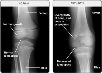
Below is an X-ray of a normal knee joint versus an arthritic knee joint. The knee on the right shows the effects of arthritis. Inflammation and increased blood flow to the joint causes bone overgrowth, decreased joint space and decreased bone density (bone appears not as white in an X-ray, which means that it is less dense. This is called osteopenia).
Why are X-rays done?
Usually, at the beginning of the disease, X-ray results are normal and therefore X-rays are not used to diagnosis JIA. X-rays are often used to exclude other problems. They can also be used to make sure the JIA has not caused early damage to the bones.

How are X-rays done?
Your child may first need to change into a hospital gown. Then they will be asked to stand next to the X-ray film. The X-ray machine will be turned on for a few seconds. During the X-ray, invisible beams of radiation will pass through your child's body to make a picture on the film. They will need to hold still for two or three seconds so the picture doesn’t blur.
Usually an X-ray is taken from the front and then the side. Your child won’t feel a thing. Also, because very little radiation is released through the X-ray, it won’t cause damage to your child's body.

