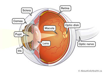
The anatomy of the eye
The eye has many parts that must work together to produce clear vision:
- The sclera, or white part of the eye, protects the eyeball.
- The pupil, or black dot at the centre of the eye, is an opening through which light can enter the eye.
- The iris, or coloured part of the eye, surrounds the pupil. It controls how much light enters the eye by changing the size of the pupil.
- The cornea, a clear window at the front of the eye, covers the iris and the pupil.
- A clear lens, located behind the pupil, acts like a camera lens by focusing light onto the retina at the back of the eye.
- The retina is a light-sensitive inner lining at the back of the eye. Ten different layers of cells work together in the retina to detect light and turn it into electrical impulses.

The retina
Special cells called cones and rods are located in the retina. These cells are known as photoreceptors and help absorb light.
Cones
The majority of the cones are located in the macula, or central area, of the retina. Cone cells help us see colour and detail. Similarly, the macula allows us to read and clearly recognize people's facial details, such as eye colour and whether they have freckles.
Rods
The majority of the rods are located in the peripheral, or outer area, of the retina. Rod cells allow us to see in poor lighting and give us our night vision.
How the eye sees
For people with normally functioning eyes, the following sequence takes place:
- Light reflects off the object we are looking at.
- Light rays enter the eye through the cornea at the front of the eye.
- The light passes through a watery fluid (aqueous humor), and enters the pupil to reach the lens.
- The lens can change in thickness to bend the light, which will focus it onto the retina at the back of the eye.
- On the way to the retina, the light passes through a thick, clear fluid called a vitreous humor. The vitreous humor fills the eyeball and helps maintain its round shape.
- The light then reaches the back of the eye and hits the retina. The retina translates the light into electrical impulses which are then carried to the brain by the optic nerve.
- Finally, the visual cortex (or centre) of the brain interprets these impulses as what we see.
What normal vision is like
To understand the vision of someone with an eye condition, it can be helpful to know what normal vision is like.
Imagine a scenario where two people are sitting on the couch in front of you. If you look directly at Person A, you are able to use your macula, or central vision, to see the details of their head and face. Maybe they have freckles, brown eyes and black hair.
At the same time, you are aware that Person B is sitting on the couch beside Person A. However, you are not able to see the same amount of detail on their face. For example, you may only see dark areas where their eyes are. To see Person B, you are using the rest of your retina or peripheral vision. Seeing clearly and sharply in the centre, and blurry in the periphery is considered normal vision.
Vision problems
A problem with any part of the eye can cause vision problems. There are many types of eye conditions that can affect vision in different ways. In some cases, the lens does not focus correctly, or the shape of the eyeball is not round, so the image will appear blurry. This can often be corrected with prescription glasses or contact lenses. When an image is focused behind the retina, this is referred to as far-sightedness. When an image is focused in front of the retina, this is referred to as near-sightedness (myopia).
Some eye conditions affect the retina. For some people, only their peripheral vision is affected, which can cause tunnel vision. For others, only their central vision is affected, which can lead to the formation of blind spots (scotomas). Finally, other eye conditions that may cause vision problems include cloudiness in the lens (cataract), increased eye pressure (glaucoma), damage to the cornea, or problems with the eye muscles.
Visual field (VF)
Visual field (VF) is the term used to describe the width of our peripheral vision. Normally, we see with a VF of 180 degrees or half a circle. When looking straight ahead, we should be able to sense motion off our right or left shoulder.
How VF is measured
Visual field can be measured in different ways. Normally, VF is measured using an automated machine called a Humphrey Visual Field Analyzer. Occasionally, a manual test called a Goldmann VF exam is used.
A person’s VF is measured by asking them to focus their eyes straight ahead. Next, a light source is brought into their field of vision from the side. The person then pushes a button when they see the light. The results of the VF test determine whether a person has tunnel vision and blind spots.
Visual fields can be measured in one eye at a time or using both eyes at once (binocular fields). A person with blind spots in both eyes may need to have a binocular visual field test to make sure that one eye sees in an area that is a blind spot in the other. This is important information when determining whether someone has a VF that meets the government standards for driving (see below).
Visual acuity (VA)
Visual acuity (VA) is defined as the clarity of the image seen by the eye. VA is tested using an eye chart at a distance of 20 feet (six metres). From testing many patients, eye doctors have determined what the average VA is for most people when standing 20 feet away from an eye chart. This measurement is called "normal" vision.
Young children who are unable to read the letters on the chart will have their vision checked using preferential looking tests where cards with lines or pictures are held up for a child to look at. The child does not need to verbally respond but will often look to the side (left or right, up or down) of the card with the picture, which will indicate the child can see it. The cards will get progressively more difficult to see, and the test continues until the child stops responding. The cards are graded and the level of vision on the 20/20 scale can be estimated.
Normal vision is 20/20 VA. In metric units, it is called 6/6 VA because 20 feet is equal to six metres. A VA of 20/20 means that when standing 20 feet away from the eye chart, a normal sighted person can see line 20 clearly.
A person with poor VA may be able to use corrective lenses (glasses or contact lenses) to correct their vision to 20/20. If corrective lenses cannot correct vision to 20/20, or at least 20/50, then the person's vision is considered too blurry to drive a car.
Examples of visual acuity
A VA of 20/50 means that a person standing 20 feet away from the eye chart will see what a 20/20 sighted person can see when standing 50 feet away. So, a person with 20/50 vision would need to stand 30 feet closer than a 20/20 sighted person to see the same amount of detail.
A person with a VA of 20/200 is able to read the biggest letter on an eye chart (usually the letter E), but all the other lines on the chart are blurry. A VA of 20/200 means that a person needs to be 20 feet from an eye chart to see the same amount of detail that a 20/20 sighted person can see from 200 feet away.
It should be noted that 20/20 VA does not always mean perfect vision. It is average vision. There are other vision skills that contribute to overall visual ability. These include:
- peripheral awareness or side vision
- eye co-ordination
- depth perception
- ability to focus
- colour vision
Definitions of "legally blind"
The term “legally blind” has a few different meanings. It is important that you ask your optometrist or ophthalmologist what the standard definition of “legally blind” is in your province, state or country.
Driving
To drive legally with a private G class license in Ontario, a person must have a VA of at least 20/50, with both eyes open with corrective lenses. The person's VF must be at least 120 degrees and not interrupted by blind spots. The VF must also be at least 15 degrees above and below the fixation point with both eyes open.
A person can have their vision classified as “legally blind” for driving when their VA or VF does not meet the guidelines set by the Ministry of Transportation (MOT). An ophthalmologist is required by law to report any patient that does not meet the MOT guidelines.
Assistive devices program (ADP)
To qualify for funding for low vision aids through the Assistive Devices Program (ADP) in Ontario, a person must have a VA of 20/70 or worse with their best correction. Such a person is classified as "legally blind for assistive devices." These people should be seen by a low vision specialist, or licensed doctor of optometry, to learn about which low vision aids are available and appropriate for their vision problems. Low vision aids include hand-held magnifiers, magnification in reading glasses and telescopes.
Disability tax credit and Registered Disability Savings Plan
To be eligible for the Disability Tax Credit Certificate in Canada, a person must have a VA of 20/200 or worse or a VF of less than 20 degrees. The Disability Tax Credit Certificate (form T2201) is available at www.cra-arc.gc.ca. If eligible, a person is entitled to a Registered Disability Savings Plan (RDSP). With the RDSP, the government matches your contribution up to a maximum of $3,000. Ask your bank for details.